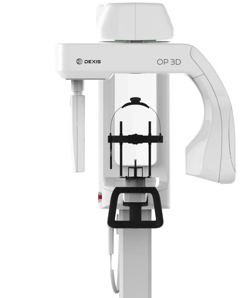
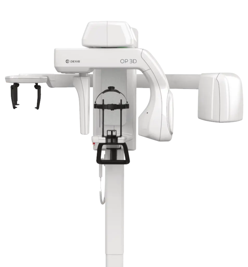
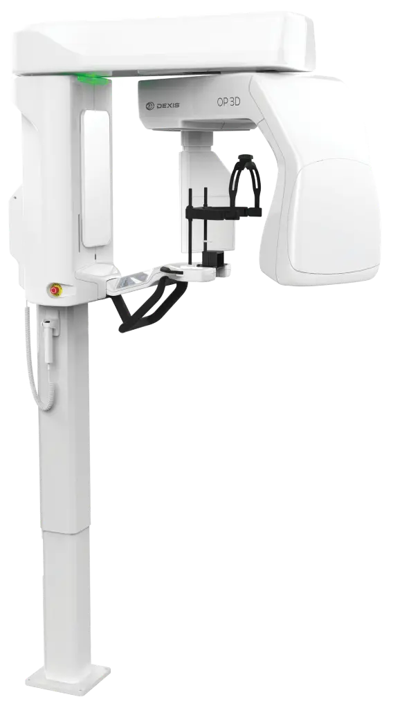
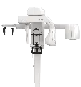
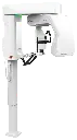
Dexis ORTHOPANTOMOGRAPH OP 3D
The DEXIS OP 3D is a unit designed for advanced dental imaging needs. It is a complete X-ray platform that provides easy-to-use features throughout the entire dental imaging workflow. The versatile panoramic, cephalometric and 3D programs offer image excellence for a variety of users ranging from general dental practitioners to maxillofacial surgeons. It respects the legacy of the ORTHOPANTOMOGRAPH product family while renewing the meaning of image quality, efficiency and ease-of-use. The OP 3D imaging unit replaces lead typically used for tubehead radiation shielding designs with a more ecological and environmentally friendly alternative that provides equivalent radiation attenuation, and its power save feature reduces overall energy consumption of the practice.
Key Features
- Fully upgradeable: choose to add cephalometric imaging, or fully upgrade to add 3D imaging
- 9-second standard and pediatric panoramic scan times, providing clear image definition with fewer movement artifacts and lower patient dose
- ORTHOfocus feature: automatically obtains the optimum panoramic image layer, enabling forgiving patient positioning and consistent image quality
- Multilayer pan: optionally supplies five layers within one exposure at the same scan time and dosage as a single panoramic exposure
- Bitewing-like view as a quick alternative to intraoral bitewing imaging
- ORTHOselect user interface: intuitive selection of imaging area (individual teeth, upper/lower jaw, or TMJ), with automatic optimum FOV
- SMARTVIEW 2.0 user-interface: two-dimensional scout images before CBCT examination, with adjustable FOV location and height, supporting ALARA radiation protocols
- QUICKcompose feature for fast preview of panoramic, cephalometric, and 3D images
- Metal Artifact Reduction (MAR), active across all FOV sizes, aiding endodontics, implants, and maxillofacial imaging
- Edge Preserving Noise Reduction (EPNR) filter, smoothing noise while preserving sharp edges and clinical detail
- Variety of resolutions: Low Dose Technology (LDT), Standard, High resolution, and Endo resolution (80 μm voxel, available for 5x5 FOV)
- ORTHOceph Plus design (cephalometric): patented design keeps correct height for CEPH imaging after a panoramic scan; dedicated X-ray source for high capacity and minimal patient dose
- Cephalometric protocols: lateral and pediatric lateral projections (adjustable field width), posterior-anterior (PA) projections, carpus imaging (optional holder)
- Easy wheelchair accessibility
- Supports implantology, endodontics, impactions, TMJ, trauma, and periodontics
Technical Data
| Specification | Value |
| 2D Panoramic | |
| Image receptor | CMOS |
| Pixel size (sensor & image) | 99 μm |
| Tube voltage | 60 - 90 kV |
| Tube current | 2 - 16 mA |
| Scan time | 9 s |
| Image field height | 147 mm |
| Imaging programs | Standard, Segmented, Pediatric, Lateral TMJ, Bitewing |
| 2D Cephalometric | |
| Image receptor | CMOS |
| Pixel size (sensor & image) | 99 μm |
| Tube voltage | 60 - 95 kV |
| Tube current | 2 - 14 mA |
| Scan time | 10.5 and 8.1 s |
| Image field height | 180 - 223 mm |
| Image field width | 160 - 260 mm |
| Imaging programs | Lateral and Pediatric Lateral (adjustable field width), Posterior-Anterior (PA), Carpus* |
| 3D CBCT | |
| Image detector | CMOS |
| Image voxel size | 80 - 400 μm |
| Tube voltage | 95 kV |
| Tube current | 2 - 12.5 mA |
| Scan time | 10 - 20 s |
| Image volume sizes (H x Ø) | 5x5, 6x9, 9x11, 9x14 cm (optional); volume height/location adjustable via SMARTVIEW 2.0 |
| Others | |
| Tube focal spot | 0.5 IEC 336 (IEC 60336/2005) |
| DICOM support | Available as a software option |