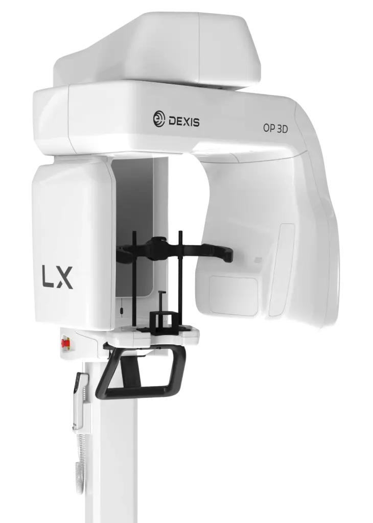
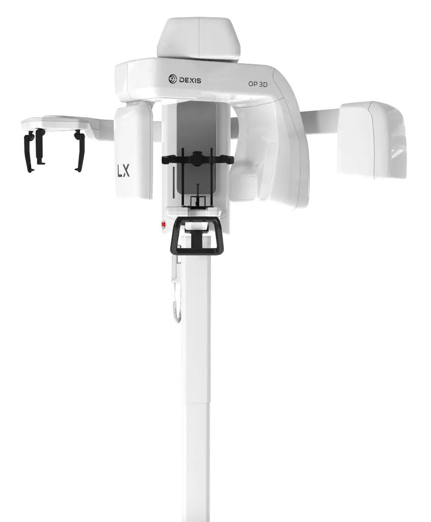
Dexis ORTHOPANTOMOGRAPH OP 3D LX
Built on OP 3D technology, the OP 3D LX is the next generation of proven DEXIS cone beam technology — a multimodality imaging platform that expands 3D diagnostic capabilities with a wide range of clinical applications supporting evolving practices and enhanced diagnostic confidence. The 2D and 3D imaging options cover a wide range of dental extraoral needs, from endodontics to complex surgical cases. This next-generation system offers flexible FOV options ranging from 5x5 up to 15x20 — the largest view option available on a DEXIS OP 3D platform to date. With short scan times, the OP 3D LX captures the maxillofacial complex and large diagnostic areas in one non-stitched scan for fast workflows.
Key Features
- Flexible FOV, from 5x5 up to 15x20 complete high-resolution scans, with 96 additional customizable options and four resolutions (including endo mode and low dose mode)
- Scalable solution: standard panoramic, extraoral bitewing and 3D imaging up to 10x10, upgradable to 12x15 and 15x20 volume sizes or cephalometric modality
- Consistent high-quality images via implant planning tools and noise reduction filters minimizing artifacts and noise
- Intuitive user interface for patient positioning, 3D/panoramic/cephalometric area selection, and instant X-ray preview without opening viewing software
- Stitch-free scans: single-pass capture with no stitching on every scan size
- Re-engineered head support, optimized for orthodontic and surgical applications without interfering with patient's soft tissue profile
- Next-generation automated ICE (Implant Contrast Enhancer) and MAR (Metal Artifact Reduction) for greater visibility of internal metal structures
- New ergonomic exposure switch with light indicator (ready/exposing) and adjustable device/remote sounds
- AI-assisted workflow features in DTX Studio Clinic: automatic focal trough, tooth-centric navigation, patient positioning correction, auto 3D tooth positioning, mandibular nerve canal annotation, merge of CBCT and intraoral scans
- Automatic Dose Control (ADC): automatically measures patient anatomy and sets exposure values for optimal image quality and dose
- Supports endodontics, maxillofacial surgery, orthodontics, TMJ analysis, airway analysis, periodontics, prosthodontics, and implant placement (single unit, All-on-4®, zygomatic)
- Optional 10-year warranty coverage
- Wheelchair accessible
Technical Data
| Specification | Value |
| Focal spot | 0.5 (IEC 60336/2020) |
| Tube voltage | 60 - 95 kV |
| Tube current | 2 - 16 mA |
| HU capacity | 35 kJ, 49,000 HU |
| Minimum total filtration | 3.4 mm Al @ 95 kV |
| Wheelchair accessible | Yes |
| Min. room height | 2100 - 2455 mm |
| 2D Panoramic | |
| Image detector | IGZO TFT |
| Sensor/image pixel size | 95 μm |
| Scan/exposure time | 1.4 - 15.0 s (Standard Pan 9.0 s) |
| Image field height | 128.4 - 187.0 mm |
| Imaging programs | Standard, HD Pan, segmented standard, pediatric, segmented pediatric, bitewing, lateral TMJ |
| Weight | 120 kg / 265 lbs |
| 2D Cephalometric | |
| Image detector | IGZO TFT |
| Sensor/image pixel size | 95 μm |
| Scan/exposure time | 6.9 - 11.3 s |
| Image field height | 180.0 - 235.9 mm |
| Imaging programs | Lateral, pediatric lateral, PA, carpus |
| Weight | 155 kg / 342 lbs |
| 3D CBCT | |
| Image detector | IGZO TFT |
| Image voxel size | 80 - 400 μm |
| Scan time | 8 - 30 s |
| Exposure time | 0.9 - 19.4 s |
| Image volume sizes (HxD) | 50x50, 60x90, 80x80, 100x100, 120x150, 150x200 mm |
| DICOM support | Yes |
| Unit dimensions | |
| Width | 1345 mm / 53.0 in |
| Depth | 530 mm / 20.9 in |
| Length | 1100 mm / 43.3 in |